This site uses cookies to improve your experience. To help us insure we adhere to various privacy regulations, please select your country/region of residence. If you do not select a country, we will assume you are from the United States. Select your Cookie Settings or view our Privacy Policy and Terms of Use.
Cookie Settings
Cookies and similar technologies are used on this website for proper function of the website, for tracking performance analytics and for marketing purposes. We and some of our third-party providers may use cookie data for various purposes. Please review the cookie settings below and choose your preference.
Used for the proper function of the website
Used for monitoring website traffic and interactions
Cookie Settings
Cookies and similar technologies are used on this website for proper function of the website, for tracking performance analytics and for marketing purposes. We and some of our third-party providers may use cookie data for various purposes. Please review the cookie settings below and choose your preference.
Strictly Necessary: Used for the proper function of the website
Performance/Analytics: Used for monitoring website traffic and interactions
Western blot analysis of rat liver lysates and microsomes confirmed higher rCYP3A1 proteinexpression in untreated SLCO2B1 +/+ compared to Slco2b1 -/ - rats.
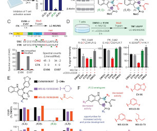
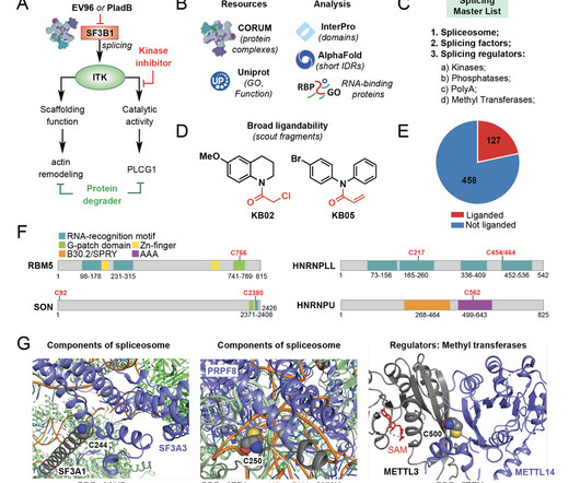
We further introduce a comprehensive list of proteins involved in splicing and leverage both cysteine- and protein-directed activity-based protein profiling (ABPP) data with electrophilic scout fragments to demonstrate covalent ligandability for many classes of splicing factors and splicing regulators in primary human T cells.
We further introduce a comprehensive list of proteins involved in splicing and leverage both cysteine- and protein-directed activity-based protein profiling (ABPP) data with electrophilic scout fragments to demonstrate covalent ligandability for many classes of splicing factors and splicing regulators in primary human T cells.
Remimazolam induced PDPK1 and p-AKT proteinexpressions, and suppressed NLRP3 proteinexpression in lung tissue of mice model. In vitro model, Remimazolam also induced PDPK1 and p-AKT proteinexpressions, and suppressed NLRP3 proteinexpression. Remimazolam interlinked PDPK1 protein.
The mRNA or proteinexpressions of examined genes were measured by quantitative real-time polymerase chain reaction or Western blot. The viability, migration, and invasion of TC cells were detected by cell counting kit-8, wound healing, and transwell assays.
Biochemical kits was utilized for checking the ATP, mitochondrial DNA, MDA, GSH, and Fe 2+ levels in the Huh7 cells, and western blot for measuring the ferroptosis and AMPK/mTOR related-proteinexpression levels.
Western blot analysis was applied to detect the proteinexpression of apoptosis HN1. The effects of matrine on tumor growth, proteinexpression of HN1, and apoptosis in vivo were validated by xenograft tumor models and histology. HN1 overexpression perceptibly reversed the above-mentioned additive effect in vitro.
In an earlier blog article, we discussed the issue of protein aggregation, how to detect it and how we screen solution conditions to overcome it. Many of the other issues with ill-behaved proteins.
Sorafenib and glimepiride simultaneously downregulated c-Maf proteinexpression to induce G1 phase arrest and apoptosis in myeloma cells. Moreover, both compounds simultaneously downregulated c-Maf proteinexpression to induce G1 phase arrest and apoptosis in myeloma cells.
OVCAR-3 and SKOV-3 cells were treated with diosgenin, cellular viability was assessed by MTT assay and apoptosis was measured by ELISA and evaluated the proteinexpression levels of apoptotic markers through western blotting. The proteinexpression levels of main components of PI3K signaling were evaluated via western blotting.
Curcumin modulates the gene and proteinexpression levels of ferroptosis mediators via JNK signaling. In addition, curcumin modulated the mRNA and proteinexpression levels of ferroptosis-related proteins including ACSL4, GPx4 and FTH1 and suppression of JNK signaling.
The results showed that hsa-miR-503-5p showed high expression, while its target gene CTDSPL presented decreased expression in LUAD. Hsa-miR-503-5p also had high expression in cisplatin-resistant LUAD cells. Binding relationship between the two genes was verified by dual-luciferase reporter assay.
Inflammatory factors in the serum were measured using ELISA assays, immunohistochemistry and qRT-PCR were performed to determine macrophage numbers and the expression of iNOS, CD86, Arg-1, and Mrc1 in myocardial tissue. The proteinexpression levels of TLR4, Myd88, and NF-κB in myocardial tissue were assessed through western blot analysis.
The proteinexpression levels of nuclear factor-erythroid 2-related factor 2 (Nrf2), matrix metalloproteinase (MMP)-2, and MMP-9 were detected by western blotting. Besides, the decreased Nrf2 expression reduced the proteinexpression levels of MMP-2 and MMP-9 ( p < .05).
Western blotting was utilized for proteinexpression and epigenetic studies utilized chromatin immunoprecipitation methods. Furthermore, KET restored acetylated histone occupancy at the Bdnf promoter IV and induced BDNF proteinexpression in DFP rats. mg/kg s.c., At 6-m following DFP exposure, KET (10 mg/kg, i.p.)
At the same time, CBG and EAA did not modify the expression/structure of proteins in relation to the non-irradiated control keratinocytes in the case of an unaccompanied use, or slightly modified the protein profile when used in a mixture.
Western blot and molecular docking indicated that compound 22 may exert antioxidant activity by activating Nrf2 proteinexpression. This might be due to the introduction of 2, 5-difluorobenzene sulfonate group in PF, which helps in scavenging free radicals under oxidative stress.
SIRT1 expression was enhanced by Icariin, and its knockdown suppressed Icariin-induced BMSC osteogenic differentiation. Moreover, deubiquitinating enzyme USP47 could stabilize SIRT1 proteinexpression.
The results showed that CA-COS inhibited nitric oxide (NO) production and downregulated the gene expression of nitric oxide synthase (iNOS), and cytokines such as tumor necrosis factor-alpha (TNF-α), IL-1β, and IL-6 without cytotoxic effect.
For instance, a dataset generated to study proteinexpression in one context might also reveal valuable information about other biological pathways or processes. Additionally, exploratory analyses can be valuable for identifying new biological markers or hypotheses.
We hypothesized that ligustrazine could protect liver by decreasing the inflammation response, protein production, and apoptosis in THS rats. The proteinexpressions were detected via western blot. Ligustrazine at doses of 100 and 1000 μg/mL was administrated in Kupffer cells isolated from THS rats.
Further, the study detected the effect of SA on cell apoptosis, lipid peroxidation, Fe 2+ level, and ferroptosis-related proteinsexpression. Finally, the effect of HMGB1 expression on SA in H/R stimulation was studied.
Linagliptin administered to the type 1 diabetic mouse heart significantly reduced the expression levels of the total and cleaved forms of ATF6, ATF4, and p-JNK, caspase 3. According to ELISA findings, TUDCA was more effective in reducing NOX 1 and MDA levels than linagliptin.
The mRNA and proteinexpression levels of p53, p-glycoprotein (P-gp), ATM, ATR, CHK1, and CHK2 were assessed through qRT-PCR and western blotting. OECM-1 and OECM-1/PTX were transfected with miR-34 mimic and inhibitor. Cellular proliferation and apoptosis were evaluated through MTT assay and flow cytometry, respectively.
RT-qPCR and western blotting were used to test the mRNA and proteinexpression levels of IL-17 and retinoid-related orphan receptor-γt (RORγt). PGE2 was highly expressed in the DE mouse model. The mRNA and protein levels of IL-17 and the key Th17 transcription factor RORγt were increased in tissues of the DE mice.
Colonic pathological changes were analyzed by hematoxylin–eosin staining, and inflammatory factor expressions in serum were determined by enzyme-linked immunosorbent assay. Immunohistochemistry and western blot were performed to quantify ferroptosis-related proteinexpressions.
Hederagenin attenuated HG-induced increase in mRNA and proteinexpression of NLRP3, ASC, and IL-1β. The secretion levels of fibrosis-related biomarkers were analyzed by ELISA. Results showed that hederagenin reduced HG-induced proliferation increase in HRMCs and HRPTEpiCs. IV, PAI-1, and TGF-β1.
In addition, SAS treatment caused a significant decrease in the mRNA and proteinexpression of xCT and GPX4, and a significant increase in ACSL4 expression in TE-1 cells treated with SAS. Flow cytometry results showed that the ferroptosis level was significantly increased after SAS treatment.
In MDA-MB-231 cell xenografts, NOSH-ASA reduced tumor size markedly, which was associated with reduced proliferation (decreased PCNA expression), induction of apoptosis (increased TUNEL positive cells), and increased ROS, while NF-kB and FoxM1 that were high in untreated xenografts were significantly reduced.
A label-free proteomics technology was employed to investigate alterations in proteinexpression in LoVo cells treated with plumbagin. Cell cycle analysis and cell apoptosis analysis were conducted to break down the anticancer impact of plumbagin on LoVo cells.
Also, Vinp downregulated α-Syn proteinexpression and MDA level, while upregulated SOD activity in the striatum of PD rats. Vinp treatment increased the horizontal movement frequency and number of squares crossed, reduced the contact time, and rotation frequency in PD rats.
The mRNA or proteinexpressions of examined genes in microglia and brain tissues were detected by quantitative real-time polymerase chain reaction or western blot.
BoNT/A (1 nM) attenuated LPS/ATP-stimulated inhibition of viability and CAMP expression and upregulation of inflammatory mediators, pyroptosis-related proteins, and ELANE expression in rat DRG neurons, which was counteracted by CAMP silencing.
Elisa's result showed that CQQNC can significantly decrease the expression levels of cPLA2, sPLA2, PGE2, cAMP, and 15-PGDH after stimulating IL-1β to bEnd.3 3 cells in a dose-dependent manner ( p < .01, 01, p < .001, 001, p < .0001). 3 ( p < .0001, 0001, p < .001, 001, p < .01).
Together, these attributes provide a strong foundation for proteinexpression with enough adaptability to produce much of the commercial and therapeutic protein market.
The expression of miR-181a-3p was inhibited; however, SHQ1 expression was increased by β-sitosterol treatment of A549/anlotinib cells. The inhibition of SHQ1, ATF6, and GRP78 proteinexpression by β-sitosterol in A549/anlotinib cells was rescued by increased miR-181a-3p.
In the analysis of network pharmacology, 269 pharmacological targets of SP, 449 pharmacological targets of FC, and 2569 targets of HS-related diseases were screened from the databases.
μM), significantly and dose-dependently induced apoptosis of SCC-12 and SK-MEL-28 cells, as evidenced by the suppression of Bcl-2 and upregulation of Bax, cleaved caspase-3, caspase-9, and PARP proteinexpression levels. The most active compounds 11 (A431: IC 50 = 5.0 μM, μM, SCC-12: IC 50 = 2.9 μM, μM, SKMEL-28: IC 50 = 4.9 μM,
Comparable Results: Achieve cell growth and proteinexpression levels similar to those obtained with smaller-scale shake flasks. Enhanced Cellular Environment: Learn about the unique design of our Nalgene flask that minimizes damaging shear stress, fostering a gentle environment for mammalian cells. Register now to secure your spot!
Etta Biotech”), to set up a high titer transient proteinexpression platform for high quality protein production using JS Bio’s transient transfection media. JS Bio becomes the exclusive cell culture supplier for Etta Biotech’s transient transfection high titer proteinexpression platform.
Predictive models for biology will span all scales, from individual proteins to the culture conditions in which cells grow. Large-scale experiments suggest that <50% of bacterial proteins and <15% of non-bacterial proteinsexpress within E. coli , yeast, and other types of organisms.
We organize all of the trending information in your field so you don't have to. Join 15,000+ users and stay up to date on the latest articles your peers are reading.
You know about us, now we want to get to know you!
Let's personalize your content
Let's get even more personalized
We recognize your account from another site in our network, please click 'Send Email' below to continue with verifying your account and setting a password.


Let's personalize your content